Preliminary paleohistological observations of the StW 573 (‘Little Foot’) skull
- Department of Archaeology, University of Cambridge, United Kingdom
- School of Geography, Archaeology and Environmental Studies, University of the Witwatersrand, South Africa
- Institut Català de Paleontologia Miquel Crusafont, Universitat Autònoma de Barcelona, Spain
- Department of Anatomy, University of Pretoria, South Africa
- Diamond Light Source Ltd, Harwell Science and Innovation Campus, United Kingdom
- STFC-Rutherford Appleton Laboratory, ISIS Facility, United Kingdom
- Core Research Laboratories, Natural History Museum, Cromwell Rd, South Kensington, United Kingdom
- Evolutionary Studies Institute, University of the Witwatersrand, South Africa
Figures
Figure 1
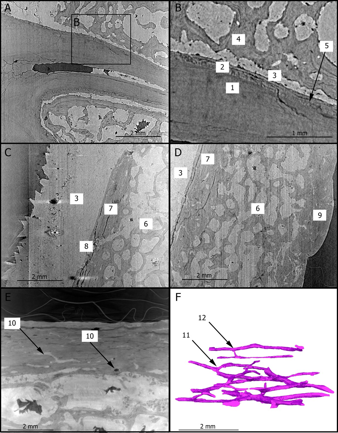
2D sections of the roots of the upper right first molar (A,B), of the cranial vault (C,D), and of the mandibular symphysis (E) of StW 573.
The close up (B) of the roots of the upper right third premolar shows the dentine–cementum junction. The Haversian canals of the mandibular symphysis is reconstructed in 3D (F) in the same orientation as in (E). 1: dentine; 2: cementum; 3: calcite; 4: trabecular bone; 5: incremental line in the cementum; 6: diploic bone; 7: inner table; 8: diploic channel; 9: outer table; 10: Haversian canals; 11: dichotomous branching; 12: transverse connection.
Figure 2
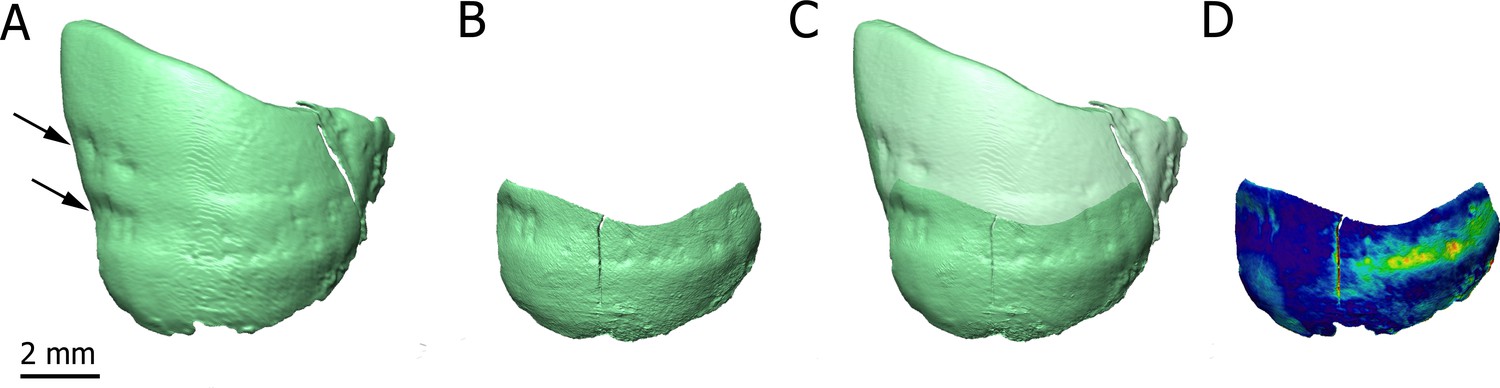
3D renderings of the buccal aspect of the enamel surface of the lower left canine of StW 573 using synchrotron-based data sets at 21.23 (A) and 7.91 µm (B).
The black arrows (A) point to enamel defects. 3D reconstructions are superimposed (C) and the distances between them are rendered by a pseudo-color scale ranging from dark blue (lowest values) to red (highest values).
Download links
A two-part list of links to download the article, or parts of the article, in various formats.
Downloads (link to download the article as PDF)
Open citations (links to open the citations from this article in various online reference manager services)
Cite this article (links to download the citations from this article in formats compatible with various reference manager tools)
Preliminary paleohistological observations of the StW 573 (‘Little Foot’) skull
eLife 10:e64804.
https://doi.org/10.7554/eLife.64804


