Pain: Why sex matters
The immune mechanisms underlying hypersensitivity to pain after nerve injury are different in male and female mice.
- Eunice Kennedy Shriver National Institute on Child Health and Human Development, National Institutes of Health, United States
- Department of Biology, Johns Hopkins University, United States
Pain cautions our bodies against harmful stimuli – such as a burning flame or the pointy end of a needle – and protects us when we are injured. These stimuli are detected by sensory neurons, which transmit signals to the spinal cord and brain. Damaging these neurons can lead to persistent and chronic pain, but the mechanisms underlying this are not fully understood.
One important player in controlling pain related to nerve damage is the immune system (Calvo et al., 2012; Scholz and Woolf, 2007). Previous work showed that injured sensory neurons release a protein called CSF1 (short for colony stimulating factor 1), which activates microglia, the main immune cell type in the brain and spinal cord. In this activated state, microglia proliferate, change their form and alter their behavior.
In 2016, a group of scientists discovered that male mice became hypersensitive to touch when their microglia were activated by nerve injury or by injecting CSF1 in to the space around the spinal cord (Guan et al., 2016). However, microglia have also been shown to be sexually dimorphic, playing different roles in disease and pain in males and females (Mogil, 2020). Now, in eLife, Allan Basbaum, Anna Molovsky and colleagues from the University of California, San Francisco – including Julia Kuhn and Ilia Vainchtein as co-first authors – report that microglia and another immune cell population respond differently to pain signals in male and female mice (Kuhn et al., 2021).
To investigate the mechanisms underlying hypersensitivity to touch, the team (which includes some of the researchers involved in the 2016 study) damaged the sciatic nerves of male and female mice lacking the gene for the CSF1 protein in their sensory neurons. Pain was assessed using the Von Frey assay, where mice are placed on an elevated grate and their paws are poked with different sized filaments (Decosterd and Woolf, 2000; Shields et al., 2003). Thick filaments will evoke a pain response that causes the mouse to flinch and withdraw its paw; whereas, thinner filaments only elicit this response when mice are hypersensitive to touch.
As shown previously, male mice deficient in CSF1 were not hypersensitive to touch after nerve injury. Female mice lacking CSF1, however, still withdrew their paws when poked with thinner filaments, suggesting that the mechanism underlying hypersensitivity in females is different to males. To confirm these findings, Kuhn et al. injected CSF1 near the spinal cord and assessed pain in the absence of nerve injury. As expected based on the previous results, the male mice became hypersensitive to touch, whereas the females did not (Figure 1). Further experiments examining the genes expressed by microglia after injection of CSF1 revealed that male mice upregulated different genes compared to females, including genes associated with disease, and the activation and recruitment of immune cells.
Figure 1
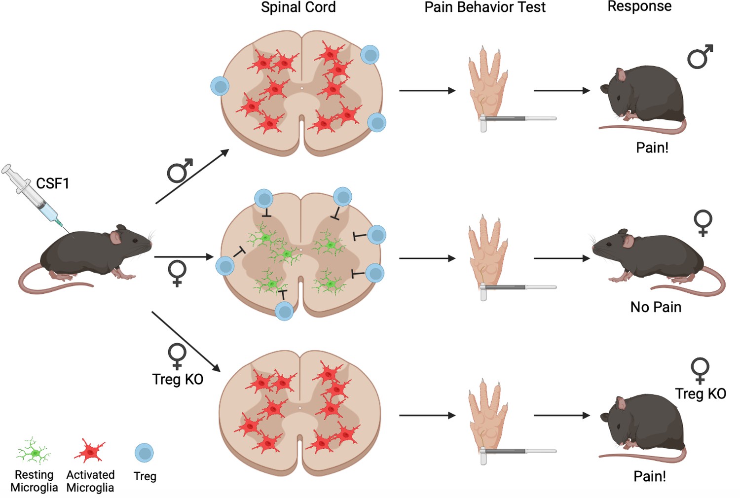
The differing effects of CSF1 injection on male and female mice.
When CSF1 is injected into wild-type mice, microglia in the spinal cord become activated (red cells) in male mice (top) but not females (middle). In females, regulatory T-cells (Tregs, blue circles) present in the membrane layers surrounding the spinal cord block CSF1 from activating microglia, which remain in the resting state (green cells); when regulatory T-cells are depleted (Treg KO; bottom), the microglia of female mice respond to CSF1 the same way as in males (bottom). During Von Frey pain assessment tests, female mice with depleted levels of regulatory T-cells and male mice exhibit the paw withdrawal response typical of hypersensitivity (top and bottom); however, female mice do not elicit a hypersensitive pain response. This indicates that regulatory T-cells suppress the activation of microglia and development of a pain response after CSF1 injection, but only in female mice.
Image credit: Figure created using Biorender.com.
Other types of immune cells are known to influence how the central nervous system works under both normal and diseased conditions. To see if any of these might be involved in female pain sensation, Kuhn et al. examined which immune cells were present in the membrane layers surrounding the spinal cords of mice injected with CSF1. Females were found to have more regulatory T-cells, which are potent inflammation suppressors. Kuhn et al. wondered if having a greater number of regulatory T-cells counteracts the effects of CSF1, so they repeated the experiments in female mice in which regulatory T-cells had been depleted. This revealed that without regulatory T-cells, female mice also develop hypersensitivity after CSF1 injection, and their microglia express a more similar pattern of genes to the microglia of males (Figure 1).
This study demonstrates that the immune system plays different roles in the pain pathways of male and female mice after nerve injury. In male mice, microglia are the major immune cell type driving pain induced by CSF1 injection, while regulatory T-cells repress this pathway in females. This work highlights the need to include males and females in scientific research, and the importance of considering sex-specific approaches for pain management. It also opens up interesting questions for future investigation. For example, it is unclear how regulatory T-cells are recruited in females after CSF1 injection, and the mechanisms underlying pain hypersensitivity in female mice remain to be discovered.
References
-
The role of the immune system in the generation of neuropathic painThe Lancet. Neurology 11:629–642.https://doi.org/10.1016/S1474-4422(12)70134-5
-
Qualitative sex differences in pain processing: emerging evidence of a biased literatureNature Reviews. Neuroscience 21:353–365.https://doi.org/10.1038/s41583-020-0310-6
-
The neuropathic pain triad: neurons, immune cells and gliaNature Neuroscience 10:1361–1368.https://doi.org/10.1038/nn1992
Article and author information
Author details
Josette J Wlaschin

Sangeetha Hareendran
Claire E Le Pichon
Publication history
Copyright
© 2021, Wlaschin et al.
This article is distributed under the terms of the Creative Commons Attribution License, which permits unrestricted use and redistribution provided that the original author and source are credited.
Metrics
-
- 1,547
- views
-
- 132
- downloads
-
- 1
- citations
Views, downloads and citations are aggregated across all versions of this paper published by eLife.
Download links
A two-part list of links to download the article, or parts of the article, in various formats.
Downloads (link to download the article as PDF)
Open citations (links to open the citations from this article in various online reference manager services)
Cite this article (links to download the citations from this article in formats compatible with various reference manager tools)
Pain: Why sex matters
eLife 10:e74935.
https://doi.org/10.7554/eLife.74935
Further reading
-
- Neuroscience
Recent studies suggest that calcitonin gene-related peptide (CGRP) neurons in the parabrachial nucleus (PBN) represent aversive information and signal a general alarm to the forebrain. If CGRP neurons serve as a true general alarm, their activation would modulate both passive nad active defensive behaviors depending on the magnitude and context of the threat. However, most prior research has focused on the role of CGRP neurons in passive freezing responses, with limited exploration of their involvement in active defensive behaviors. To address this, we examined the role of CGRP neurons in active defensive behavior using a predator-like robot programmed to chase mice. Our electrophysiological results revealed that CGRP neurons encode the intensity of aversive stimuli through variations in firing durations and amplitudes. Optogenetic activation of CGRP neurons during robot chasing elevated flight responses in both conditioning and retention tests, presumably by amplifying the perception of the threat as more imminent and dangerous. In contrast, animals with inactivated CGRP neurons exhibited reduced flight responses, even when the robot was programmed to appear highly threatening during conditioning. These findings expand the understanding of CGRP neurons in the PBN as a critical alarm system, capable of dynamically regulating active defensive behaviors by amplifying threat perception, and ensuring adaptive responses to varying levels of danger.
-
- Neuroscience
Substance-induced social behavior deficits dramatically worsen the clinical outcome of substance use disorders; yet, the underlying mechanisms remain poorly understood. Herein, we investigated the role for the corticotropin-releasing factor receptor 1 (CRF1) in the acute sociability deficits induced by morphine and the related activity of oxytocin (OXY)- and arginine-vasopressin (AVP)-expressing neurons of the paraventricular nucleus of the hypothalamus (PVN). For this purpose, we used both the CRF1 receptor-preferring antagonist compound antalarmin and the genetic mouse model of CRF1 receptor-deficiency. Antalarmin completely abolished sociability deficits induced by morphine in male, but not in female, C57BL/6J mice. Accordingly, genetic CRF1 receptor-deficiency eliminated morphine-induced sociability deficits in male mice. Ex vivo electrophysiology studies showed that antalarmin also eliminated morphine-induced firing of PVN neurons in male, but not in female, C57BL/6J mice. Likewise, genetic CRF1 receptor-deficiency reduced morphine-induced firing of PVN neurons in a CRF1 gene expression-dependent manner. The electrophysiology results consistently mirrored the behavioral results, indicating a link between morphine-induced PVN activity and sociability deficits. Interestingly, in male mice antalarmin abolished morphine-induced firing in neurons co-expressing OXY and AVP, but not in neurons expressing only AVP. In contrast, in female mice antalarmin did not affect morphine-induced firing of neurons co-expressing OXY and AVP or only OXY, indicating a selective sex-specific role for the CRF1 receptor in opiate-induced PVN OXY activity. The present findings demonstrate a major, sex-linked, role for the CRF1 receptor in sociability deficits and related brain alterations induced by morphine, suggesting new therapeutic strategy for opiate use disorders.


